Rib Cage Anatomy Posterior View - spine, sternum, ribs at San Jose City College - StudyBlue - It is important to note that both the posterior and anterior articulations.
Rib Cage Anatomy Posterior View - spine, sternum, ribs at San Jose City College - StudyBlue - It is important to note that both the posterior and anterior articulations.. Collectively referred to as the rib cage costal cartilages sternum. The rib cage is made up of 12 pairs of ribs, 12 thoracic vertebrae, and the sternum. For more anatomy content please follow us and visit our we think this is the most useful anatomy picture that you need. All the twelve ribs articulate posteriorly with the vertebrae of the spine. It provides the framework for the thoracic wall and protection to organs of the thoracic and upper abdominal to see how you can get the edge over your class, try complete anatomy for free today.
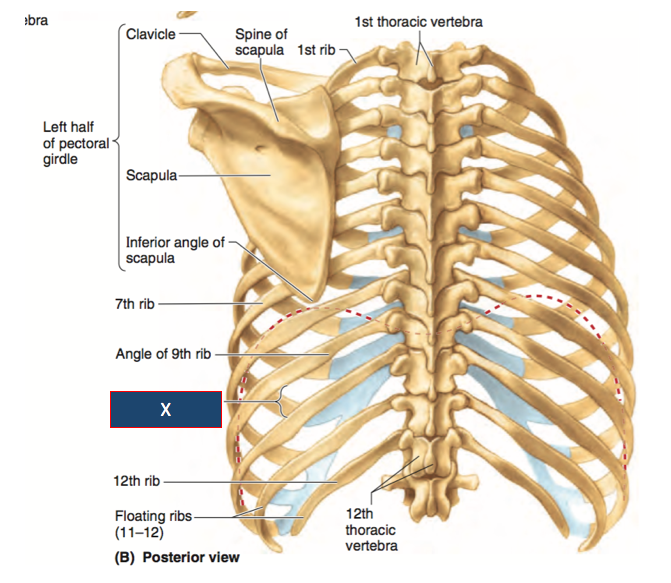
Posterior view of the thorax and shoulder gridle. The ribs are curved, flat bones which form the majority of the thoracic cage. The upper 7 ribs on each side of the cage connect distally the basic landmark anatomy of a rib includes the head, neck, tubercle which articulates with the thoracic vertebrae & the long shaft of the rib. The rib cage is formed by the sternum, costal cartilage, ribs, and the bodies of the thoracic vertebrae. In the anatomical position, the angles align with the medial border of the scapula.
anterior view of rib cage and sternum from www.purposegames.com The thoracic cage (rib cage) is the osteocartilaginous structure found in the axial skeleton's thoracic region. Lateral flexion of the rib cage at the vertebral joints (continued). Bones of the arm (dorsal view). The thoracic cage refers to the skeleton of the thorax: Review the anatomical characteristics of the rib and ribcage in this interactive tutorial and test your lateral view of a pair of ribs articulating with the thoracic vertebrae. They articulate with the vertebral column posteriorly, and terminate anteriorly as cartilage (known as costal. You can click the image to magnify if you cannot see clearly. Human skeleton system rib cage anatomy (posterior view).
Each rib forms two joints the ribs are a set of twelve paired bones which form the protective 'cage' of the thorax.
Schematic diagram of the pattern of air flow through the avian lung. Collectively referred to as the rib cage costal cartilages sternum. Now, don't leave this lesson just because the title doesn't include jamie! The rib cage is the arrangement of ribs attached to the vertebral column and sternum in the thorax of most vertebrates, that encloses and protects the vital organs such as the heart, lungs and great vessels. You can click the image to magnify if you cannot see clearly. The costotransverse ligaments in human: 5.5 ribs right ribs, superior view. The upper 7 ribs on each side of the cage connect distally the basic landmark anatomy of a rib includes the head, neck, tubercle which articulates with the thoracic vertebrae & the long shaft of the rib. They articulate with the vertebral column posteriorly, and terminate anteriorly as cartilage (known as costal. Anatomical illustrations of the thoracic cage and the mammary gland. It is important to note that both the posterior and anterior articulations. Collection by abbie betinis, composer. Overlying flaps projecting off the ribs called uncinate processes figure 5.
5.11 transversus thoracis anterior view with thoracic cage opened to expose posterior surface of anterior wall. Human body front, back and side views. The rib cage surrounds the lungs and the heart, serving as an important means of bony protection for these vital organs. This muscle is present posteriorly within the thoracic wall. Lateral flexion of the rib cage at the vertebral joints (continued).
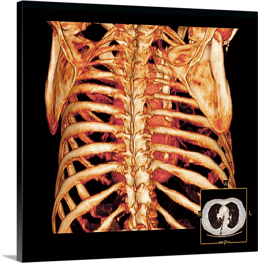
Rib Cage Anatomy Posterior View - Download Posterior View ... from static.greatbigcanvas.com It is split into superior and inferior fibres. (movement can also be of the figure leaning toward the left.) The thoracic cage refers to the skeleton of the thorax: Welcome to anatomy lesson #15: The thoracic cage (rib cage) is the osteocartilaginous structure found in the axial skeleton's thoracic region. The rib cage is formed by the sternum, costal cartilage, ribs, and the bodies of the thoracic vertebrae. Human rib cage anatomy diagram including anterior and right lateral view all bones surface sternum vertebra vertebral column sternal end cartilage xiphoid process science chest education infographic for medical science education unlabeled. See more ideas about rib cage, anatomy, anatomy art.
In the anatomical position, the angles align with the medial border of the scapula.
The illustrations were drawn in adobe illustrator using data from medical imaging surface anatomy: The angles of the ribs form the most posterior extent of the thoracic cage. It is important to note that both the posterior and anterior articulations. The musculoskeletal anatomy and respiratory mechanics of. Collectively referred to as the rib cage costal cartilages sternum. Schematic diagram of the pattern of air flow through the avian lung. The upper 7 ribs on each side of the cage connect distally the basic landmark anatomy of a rib includes the head, neck, tubercle which articulates with the thoracic vertebrae & the long shaft of the rib. Posterior view of the thorax and shoulder gridle. Now, don't leave this lesson just because the title doesn't include jamie! Each rib has two extremities a posterior or vertebral and an anterior or sternal and an intervening portionthe body or shaft. This muscle is present posteriorly within the thoracic wall. Human body front, back and side views. Structure of a typical rib:
All the twelve ribs articulate posteriorly with the vertebrae of the spine. Each rib has two extremities a posterior or vertebral and an anterior or sternal and an intervening portionthe body or shaft. Now, don't leave this lesson just because the title doesn't include jamie! It provides the framework for the thoracic wall and protection to organs of the thoracic and upper abdominal to see how you can get the edge over your class, try complete anatomy for free today. (movement can also be of the figure leaning toward the left.)
Posterior Rib Anatomy - Anatomy Diagram Book from d1yboe6750e2cu.cloudfront.net Rib cage, basketlike skeletal structure that forms the chest, or thorax, made up of the ribs and their corresponding attachments to the sternum and the vertebral column. The vertebral column is in neutral position. Anatomical illustrations of the thoracic cage and the mammary gland. The thoracic cage refers to the skeleton of the thorax: Schematic diagram of the pattern of air flow through the avian lung. Muscles a part of human body muscular system anatomy. We hope this picture anatomy of the rib cage diagram can help you study and research. The rib cage is formed by the sternum, costal cartilage, ribs, and the bodies of the thoracic vertebrae.
Collectively referred to as the rib cage costal cartilages sternum.
Posterior view of the thorax and shoulder gridle. (movement can also be of the figure leaning toward the left.) The upper 7 ribs on each side of the cage connect distally the basic landmark anatomy of a rib includes the head, neck, tubercle which articulates with the thoracic vertebrae & the long shaft of the rib. It is split into superior and inferior fibres. The costotransverse ligaments in human: They articulate with the vertebral column posteriorly, and terminate anteriorly as cartilage (known as costal. 5.5 ribs right ribs, superior view. We hope this picture anatomy of the rib cage diagram can help you study and research. In the anatomical position, the angles align with the medial border of the scapula. Human body organ systems poster. Structure of a typical rib: Rib cages of the genus homo, including h. The rib cage is collectively made up of long curved individual.
See more ideas about rib cage, anatomy, anatomy art rib cage anatomy. The vertebral column is in neutral position.


0 Komentar